
Venous Leg Ulcers
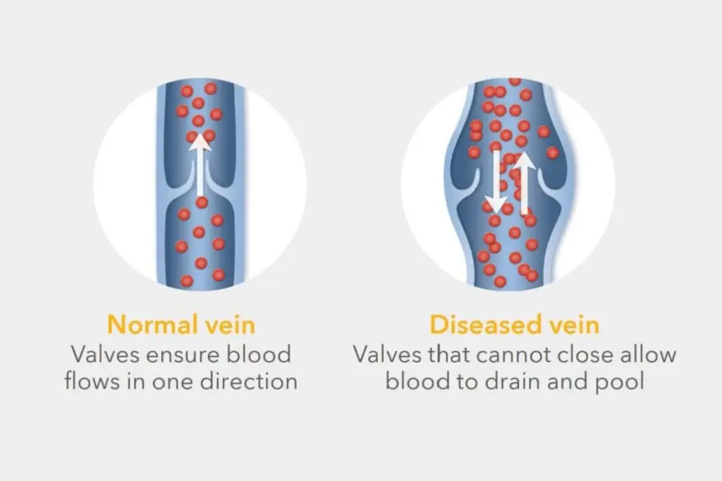
These are caused by excessive venous pressure on the skin of your lower leg. This is called chronic venous hypertension/chronic venous insufficiency(CVH/CVI). This happens due to 2 reasons : varicose veins or previous deep vein thrombosis (DVT).
Varicose Veins and Venous Leg Ulcers
Patients with varicose veins have baggy or dilated leg veins. The valves in your varicose veins become ‘loose’ and no longer work properly. Blood ‘leaks’ back (reflux) around your ankle causing swelling, pain, skin damage and ultimately leg ulcers.
Click here to read on ‘Varicose Veins’.

Stage 1: Varicose veins visible
Stage 2: Lower leg swelling, usually at the end of the day or after prolonged walking, standing or sitting.
Stage 3: Venous eczema/Varicose eczema – The skin around your ankles becomes dry, flaky and itchy.
Stage 4: Lipodermatosclerosis(LDS) & pigmentation/haemosiderosis – Leakage of blood cells under high pressure into the subcutaneous tissue(tissue beneath your skin) causes hardening of your skin and brownish discolouration on the insides of your ankles.
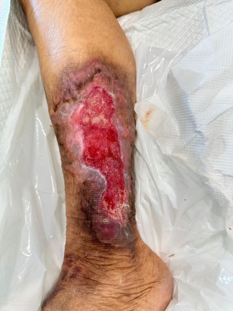
Stage 5: Venous leg ulcer – Ulceration forms from minor trauma. It does not heal despite repeated cleaning and dressings. It may produce a light yellow fluid called serous discharge. The ulcer is at risk of repeated infection.
It is important to treat varicose veins early to prevent progression of CVH skin changes and ulceration. Once your leg has progressed to ulceration, you will need urgent early RFA treatment of your varicose veins and compression bandaging to enable ulcer healing. This is supported by recent research in the New England Journal of Medicine. Click here to read about Varicose Veins Treatment.
EARLY TREATMENT OF YOUR VARICOSE VEINS PREVENTS SKIN DAMAGE AND LEG ULCERS. IT IS MORE THAN A COSMETIC CONCERN.
Stage 1 – Visible varicose veins

Stage 2 – Leg swelling
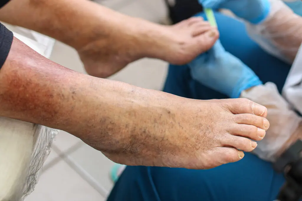
Stage 3 – Varicose eczema/dry skin
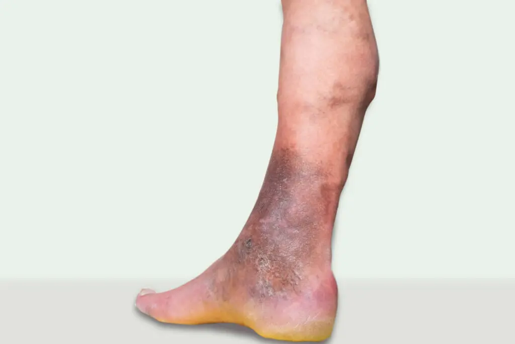
Stage 4 – Pigmentation & hardening of skin(LDS)
Stage 5 – Ulceration
Varicose Veins Bleeding
Any varicose veins is at risk of bleeding due to minor trauma. Sometimes, the bleeding is mistaken for arterial bleeding because the blood often spurts out/’shoots out’ due to the chronic venous hypertension.
First aid for varicose veins bleeding : Sit down and elevate the affected leg. Place a rolled-up gauze swab or clean kitchen towel over the bleeding varicose veins. Press firmly for at least 5 minutes. Do not be tempted to look at it until the time is up. If the bleeding has not stopped, see a doctor.
Click here to read the Dr Yow’s Instagram post on Pressure Bandaging for Bleeding.
Deep Vein Thrombosis and Venous Leg Ulcers
The legs have 2 sets of veins, a superficial and deep system which ensure healthy drainage of blood in the legs back to the heart. The deep veins are situated deep within leg muscles. In situations where they are compressed, prolonged immobility after long-haul flights, major surgery or stroke, changes in blood due to drugs, cancer or dehydration, blood clots can form in the deep veins. This is called deep vein thrombosis(DVT). DVTs can damage the valves in the deep veins.
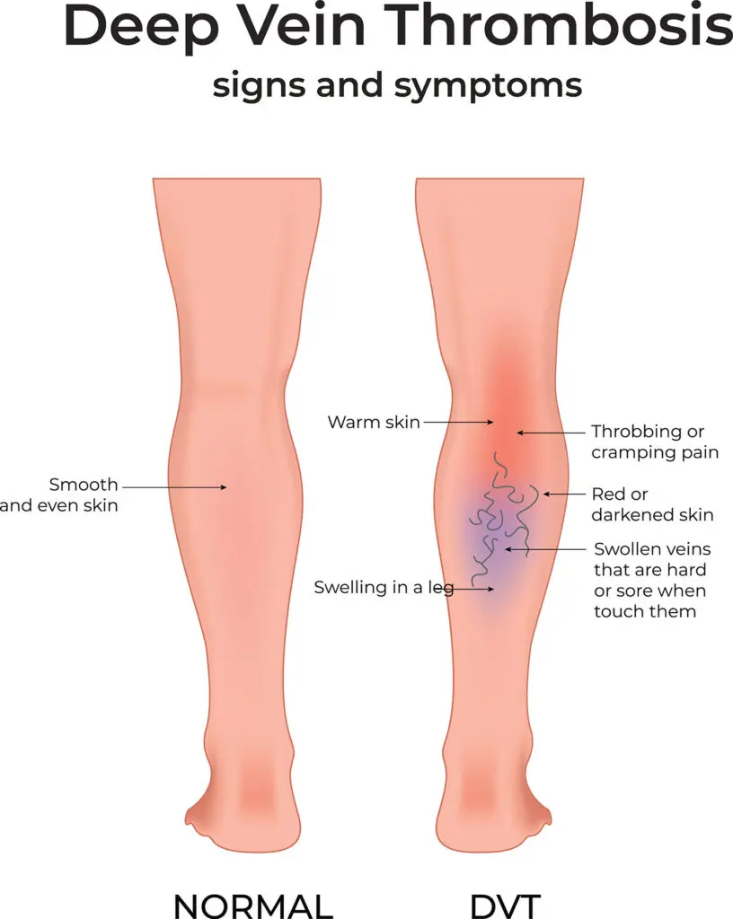
DVT symptoms & signs
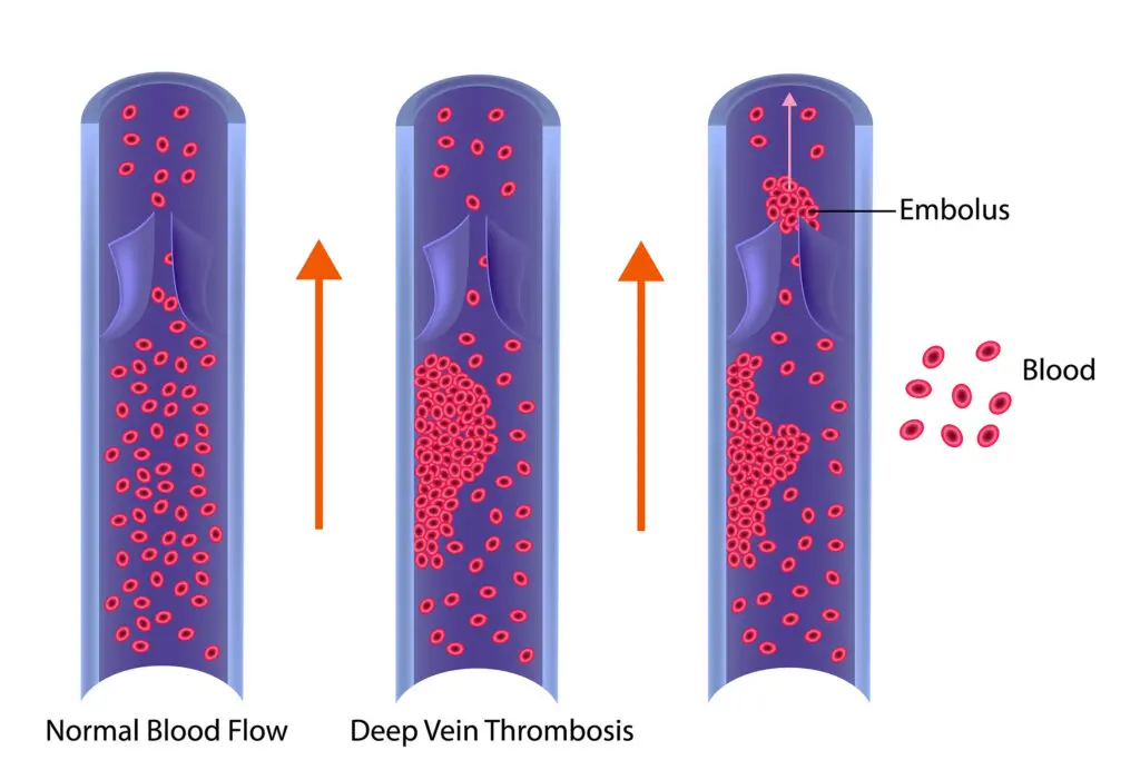
DVT & PE process
Testimonial: Jayden Lim
First and foremost, my 86-year-old grandpa has been experiencing chronic leg swelling and venous claudication since June 2023. He has been under the care of two specialist doctors at prominent hospitals in the Klang Valley.
Initially, my grandpa was referred to a consultant surgeon doctor, who advised my grandpa to meet up with a consultant general and vascular surgeon at a different hospital. Unfortunately, the general and vascular surgeon doctor did not provide any additional treatment options and merely suggested continuing the blood thinners for another month. Despite following this advice, the swelling in his leg persists.
Consequently, my uncle arranged for my grandpa to see Dr. Yow, who was recommended by a friend after my grandpa had been experiencing anxiety for nearly a year. After consulting with Dr. Yow, he expressed full confidence in managing my grandpa’s condition, despite the chronic situation of his case. Providing that grandpa’s high-risk due to his elderly, Dr. Yow’s extensive experience and expertise have reassured him that Dr. Yow is well-equipped to handle the complexities of such a high-risk patient.
Thankfully, my grandfather has made a full recovery. Therefore, I highly recommend consulting Dr. Yow for anyone dealing with post-thrombotic syndrome, as his expertise can be crucial for timely and effective treatment.
It is crystal clear that Dr. Yow is responsible and he is capable of making decisions on carrying out surgery. He has high-spirited medical ethics and moral cultivation.
I believe that Dr. Yow will emerge as a leading authority in the field of vascular medicine.
I, along with my family, would like to express my heartfelt gratitude to Dr Yow Kuan Heng and his teams in Pantai Hospital Kuala Lumpur for putting effort into treating my grandpa with care and condition.
5 Star Google Review ★★★★★
DVTs can cause problems in 2 phases : Early and late complications. The most dangerous early complications of DVTs are that the clot which forms in the deep veins of the legs can break off and travel to the lungs. This is called pulmonary embolism(PE). If the pulmonary embolus is large enough to block the circulation in the lungs and the heart, it can cause a life-threatening cardiac arrest. Traditionally, DVTs are treated with blood thinning drugs called anti-coagulants. These may be in the form of injections or tablets. Leg swelling is treated with compression stockings and elevation.
Increasingly, if there is a very large clot extending into the iliac and femoral veins above the knee, Vascular Surgeons are called upon to perform an endovascular thrombectomy. Mr Yow has performed endovascular mechanical thrombectomies/thrombolysis in patient’s with May-Thurner Syndrome(compression of the left common iliac vein by the right common iliac artery resulting in an iliac venous clot/DVT) and Paget-Schrotter Syndrome(compression of the subclavian vein of the arm from a large 1st rib/cervical rib causing a subclavian DVT). Both patients had very good outcomes.
In the late phase after DVTs, patients can develop Post-Thrombotic Syndrome(PTS). This can be a disabling condition that can cause many long-term problems. Hence, many Vascular Society guidelines now recommend early interventional treatment of high/proximal DVTs(any clot that extends into the large groin and pelvic veins) such as mechanical thrombectomy or cather directed thrombolysis mentioned above.
In chronic or long-standing PTS, the prolonged obstruction of the deep venous system and venous reflux due to an old, extensive DVT will lead to leg swelling, pain with walking and standing(venous claudication) and ultimately leg ulcers. In such situations, it may be necessary to perform a balloon angioplasty and stenting of the narrowing. Mr Yow has performed venous stenting to help relieve leg swelling and recurrent skin infections in an elderly patient.
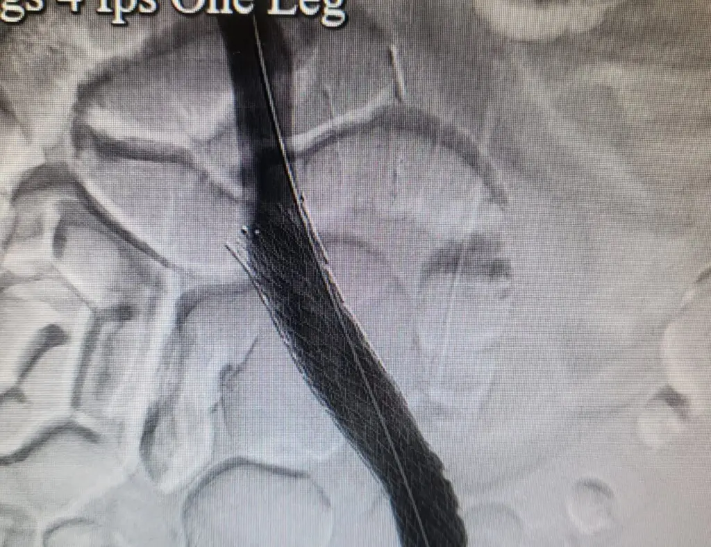
Completion angiogram of metallic stent in common iliac vein for Post-Thrombotic Syndrome
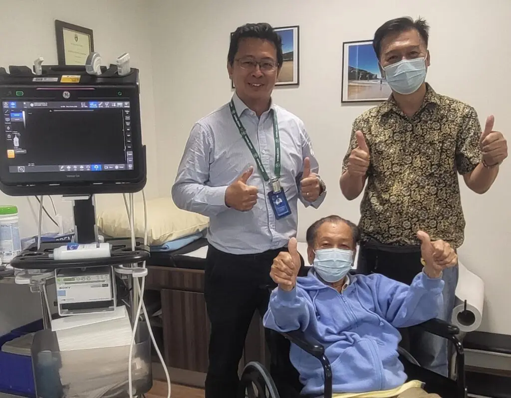
Mr Yow with elderly DVT patient who had full resolution of his leg swelling after venous stenting
Arterial Leg Ulcers
Arterial leg ulcers are caused by hardening, narrowing and blockage of your leg arteries in a disease process called atherosclerosis. In the legs, this is called peripheral arterial disease(PAD). This same process is what happens in the heart to cause angina and heart attacks. In the neck(carotid) arteries, it causes strokes. This is because the arteries throughout your body are exposed to the same cardiovascular risk factors as the blood circulates through the body. These Vascular risk factors are the chemicals in tobacco smoke(cigarettes or cigars), LDL-cholesterol, high sugars in diabetes and excessively high blood pressure. Your family history may predispose you to have one of these last three conditions. Being elderly is another risk factor as atherosclerosis is a degenerative condition.
How Do I Know I Have Peripheral Arterial Disease(PAD)?
- Pain in my calves or thighs after walking a certain distance. The pain goes away with rest.
- Severe pain/numbness in my feet or toes in the early AM which is improved by dangling the legs out of bed. The pain wakes me up from sleep. Recently, the pain is so bad I have to sleep in a chair.
- Ulcers/Black patches on my toes or the outside of my leg that do not heal.
- My feet feel cold and look blue with the above symptoms and any of the risk factors below.
- I smoke tobacco.
- I have diabetes mellitus.
- I have high blood pressure.
- I have high cholesterol.
- I have a history of coronary heart disease or strokes.
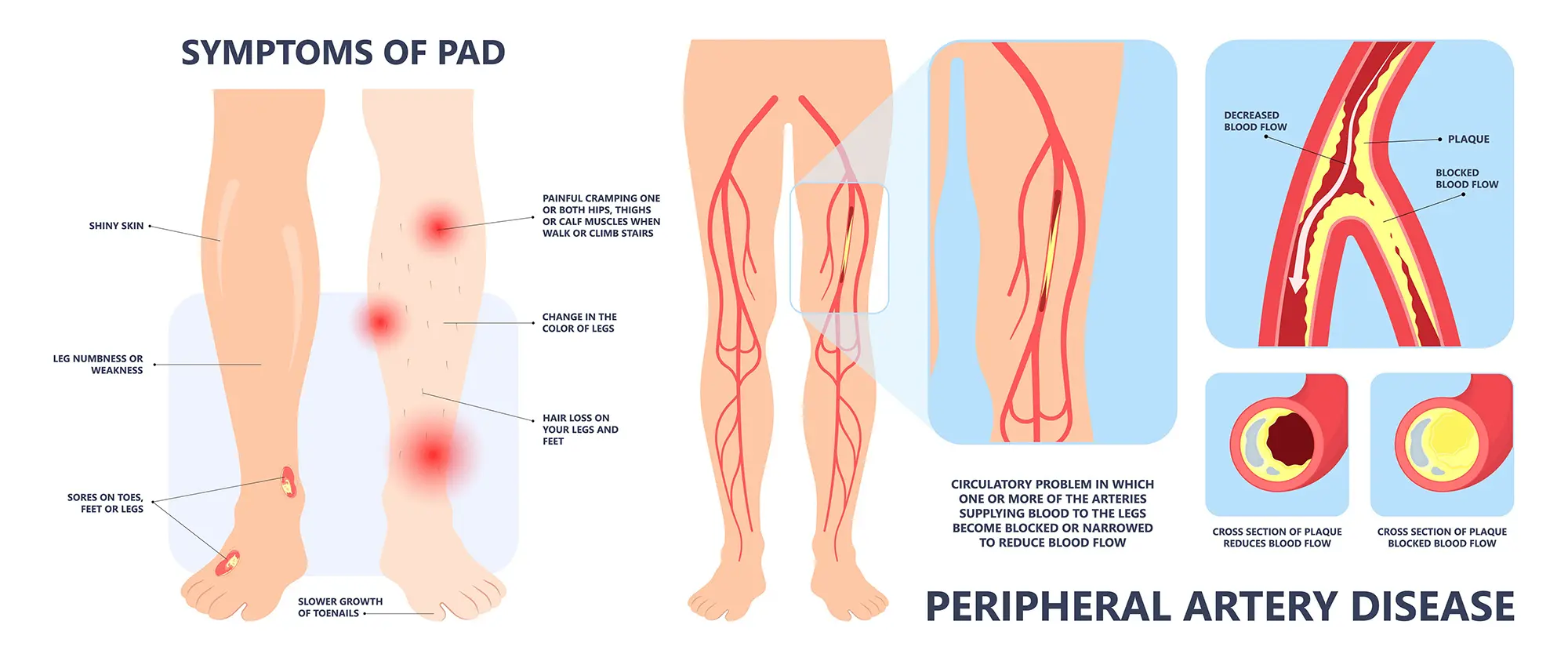
Atherosclerotic blockage of your leg arteries prevents crucial oxygenated blood from reaching your lower limbs. Any minor bump, cut or break in the dry skin of your leg produces an ulcer usually located anywhere on the exposed foot or on the outside ankle. Without crucial oxygen and important white blood cells to repair and heal the ulcer, the ulcer will get bigger over time.
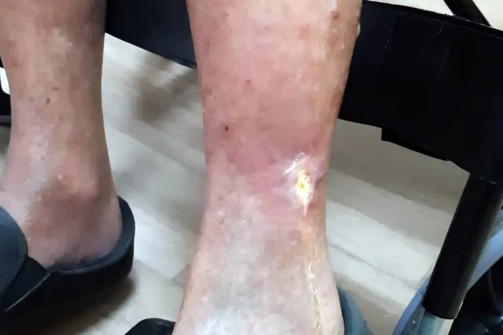
Arterial leg ulcer : Typically on outside of leg. Skin is shiny and dry due to loss of hair, sweat and sebaceous glands which keep skin healthy. The leg is more swollen than the other side as the patient has been sleeping in a chair

Arterial foot ulcer that has been neglected & enlarging. Skin around ulcer is turning black/gangrenous. Patient is at risk of amputation without Vascular intervention.
The patient often complains of night-time pain in his or her foot after being asleep for a few hours. This ischaemic rest pain can be relieved by hanging it down or over the edge of the bed. Some patients resort to sleeping in a chair. The foot is often swollen and red due to the prolonged dependency and maximally dilated blood vessels in the skin. This is called a sunset foot.(See below) Patients often liken it to an over-ripe tomato due to redness, swelling & occasional weeping fluid.
Arterial leg or foot ulcers often progress to gangrene(black toes).
IT IS VERY IMPORTANT TO SEE A VASCULAR SURGEON IMMEDIATELY. WITHOUT URGENT TREATMENT, THE PATIENT IS AT RISK OF LOSING THEIR LEG.
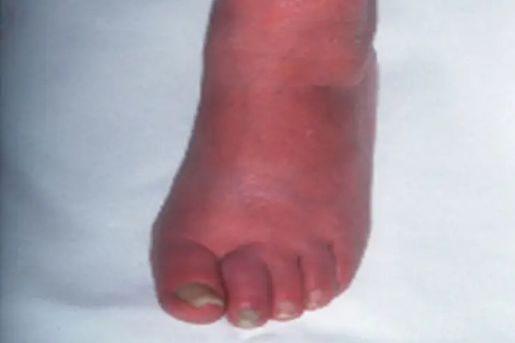
Sunset foot
At-risk foot due to peripheral arterial disease prior to ulceration and gangrene.
Foot is:
- Swollen(due to sleeping in chair) AND
- Red (foot capillaries are fully dilated due to lack of oxygen)
Arterial leg ulcers and gangrene represent the severe end/end stage of the spectrum of atherosclerotic disease in the lower limbs. This is called critical limb ischaemia (CLI) or chronic limb threatening ischaemia(CLTI). Without treatment, the patient is at risk of losing their leg i.e. amputation.
The role of the Vascular and Endovascular Surgeon is to prevent or stop amputation. We do this by treating the blockage or narrowing using open bypass, angioplasty or a hybrid of both methods.
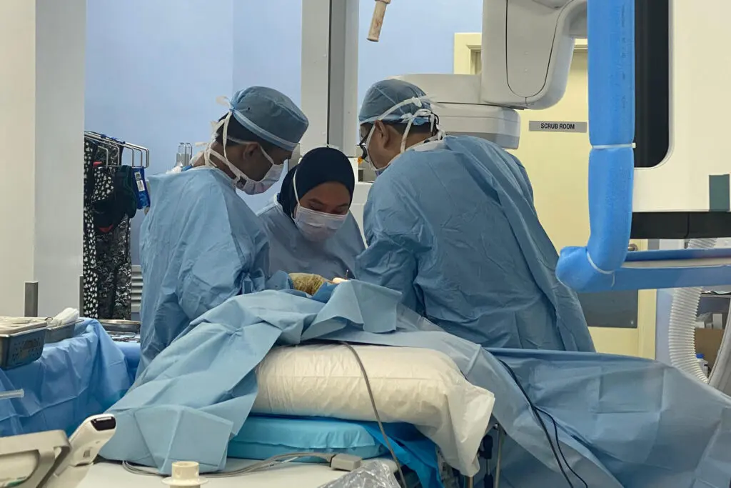
Mr Yow performing bypass surgery with his team
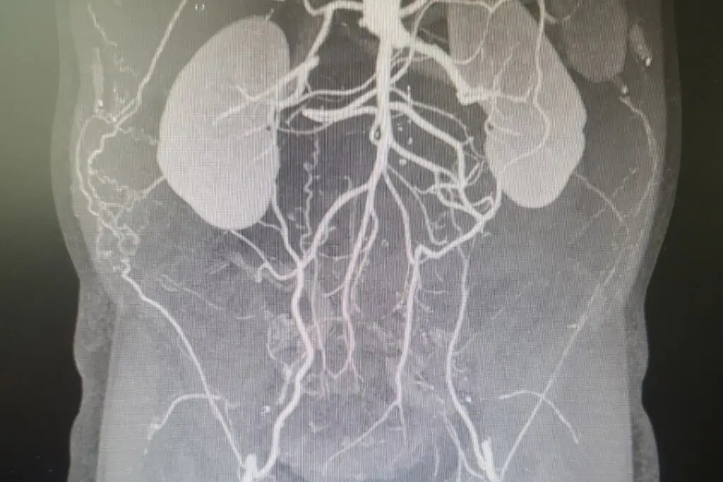
CT angiogram showing severe PAD blockage of the entire aorta from the kidneys to the femoral arteries in the groins and thighs
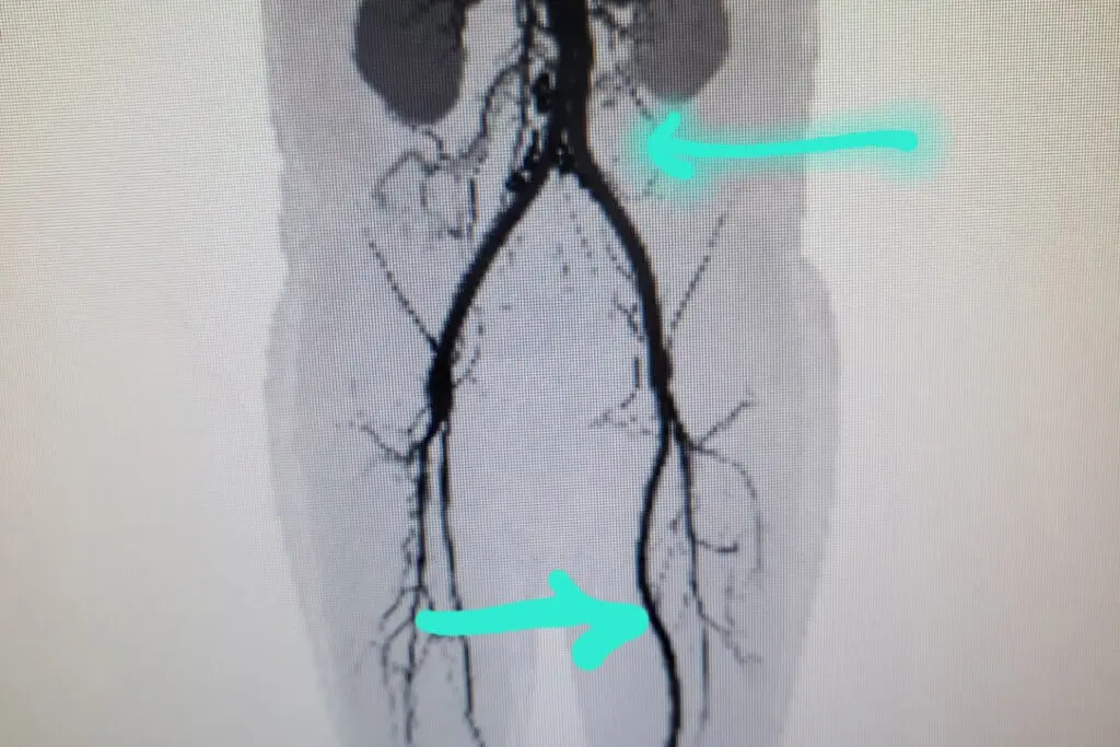
CT angiogram showing after Mr Yow performed a 2-stage operation of bypass from aorta to femoral arteries and a 2nd operation from left femoral artery to the popliteal artery
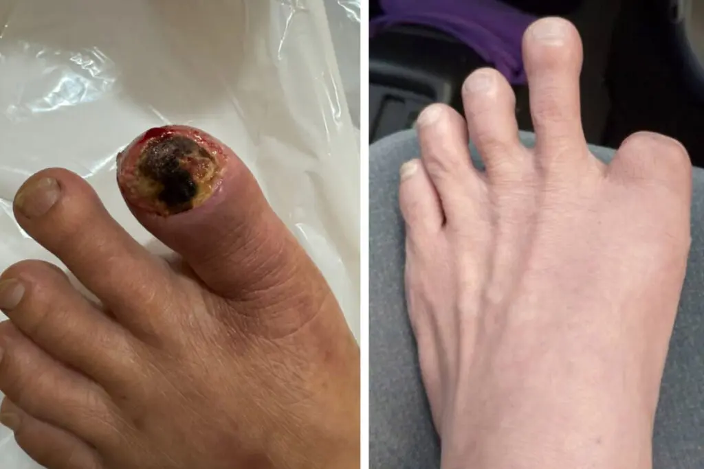
Patient was a heavy smoker who had numbness in both legs and gangrene in his left foot before Mr Yow’s double bypass. After the 2 bypass operations, he had a pink foot.
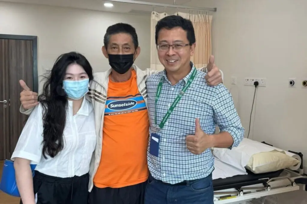
Mr Yow with the patient and his daughter 1 month after his double leg bypass. He was saved from bilateral leg amputations.
Patient Review – Jessie Tan
I and my family would like to express ours gratitude and appreciation to Dr. Yow Kuan Heng and his team.
Dr. Yow helped my brother by doing a major artery bypass surgery for more than 12 hours. The operation was successful and saved my brother’s life. Dr. Yow is also a specialist on leg ulcers and save my brother’s leg as well.
Dr. Yow is a very professional, skillful and responsible doctor. He clearly explained everything to us before surgery.
Strongly recommend Dr. Yow Kuan Heng if anyone facing vascular and leg ulcers problems.
Diabetic/Neuropathic Leg Ulcers/Mixed Arterial & Neuropathic Ulcers/Diabetic Foot
Diabetes mellitus is a complex and terrible disease. Due to the high circulating blood sugars in a diabetic patient, virtually all systems in the body are adversely affected. These include your arteries, heart, kidneys, nerves, eyes, skin and crucially your ability to fight infection. Due to the rise in sedentary lifestyles, ultra-processed foods(‘junk food’) high in sugars and obesity, Malaysia now has the highest rate of diabetes in the ASEAN region. As of 2025, more than 1 in 5 of adult Malaysians has diabetes. According to the recent Malaysian National Morbidity & Mortality study, diabetes incidence in the population is rising by 1% per year. This means 170, 000 new diabetics per year.
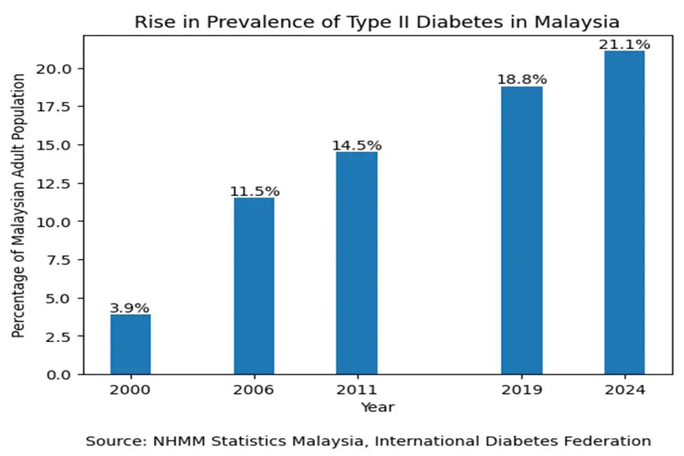
Year-on-year rise in prevalence of Type 2 Diabetes Mellitus in adult population of Malaysia. The rate of diabetes has risen more than 500% in the last 25 years. This mirrors global trends.
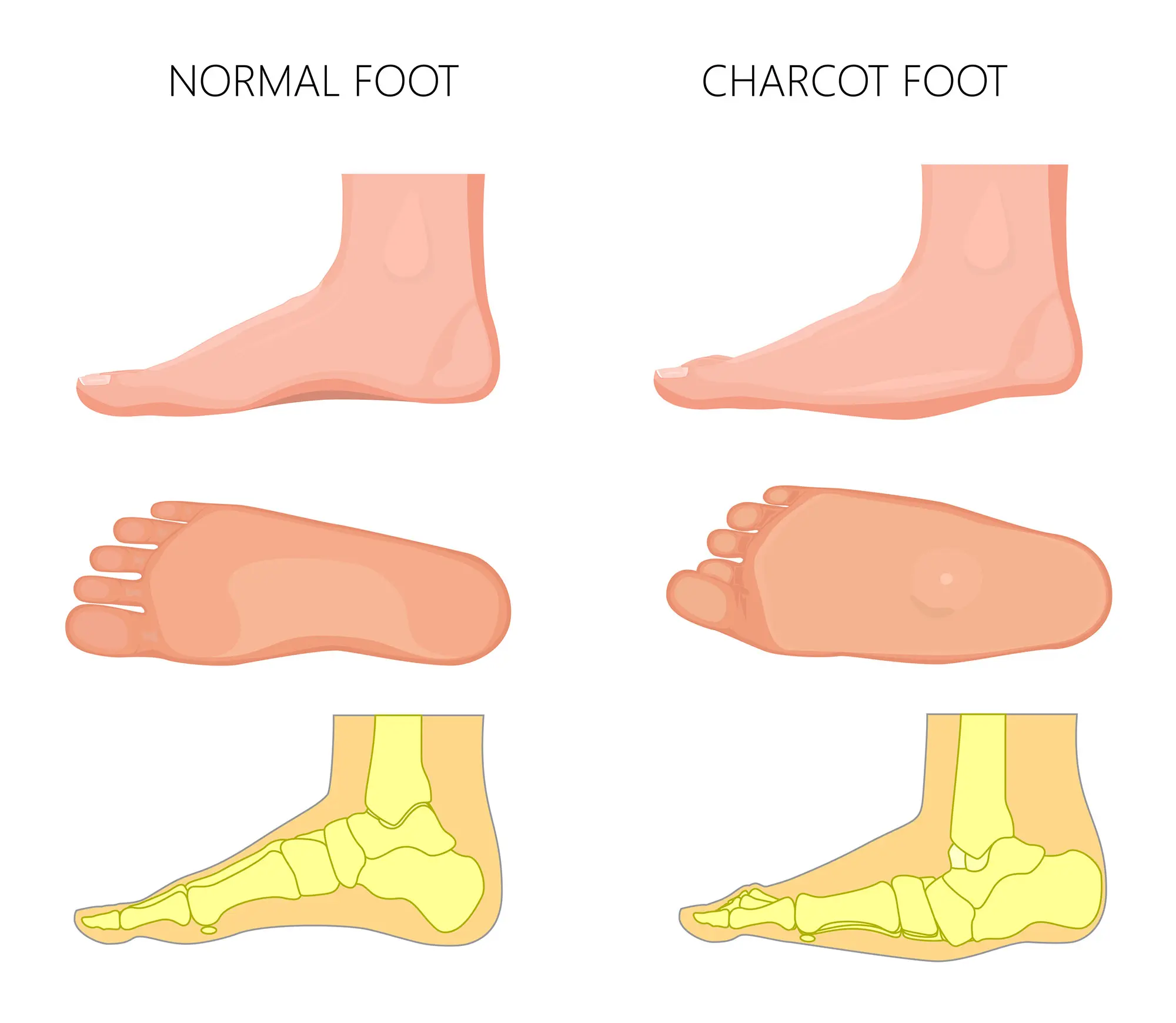
In the legs, long-standing diabetics often have damage to the nerves(neuropathy) and arteries(atherosclerosis & micro-angiopathy). Due to neuropathy, patients often cannot feel minor trauma to their feet, unlike non-diabetics. For example, patients with diabetic neuropathy often cannot feel an injury from a small piece of broken glass or excessive pressure from a tight shoe. This results in ulcers in the pressure or exposed areas of the foot. Damage to the nerves that sense joint position also cause unnecessary pressure on some foot joints more than others, resulting in foot deformity. These deformities (Charcot’s foot) increase the risk of pressure ulcers.
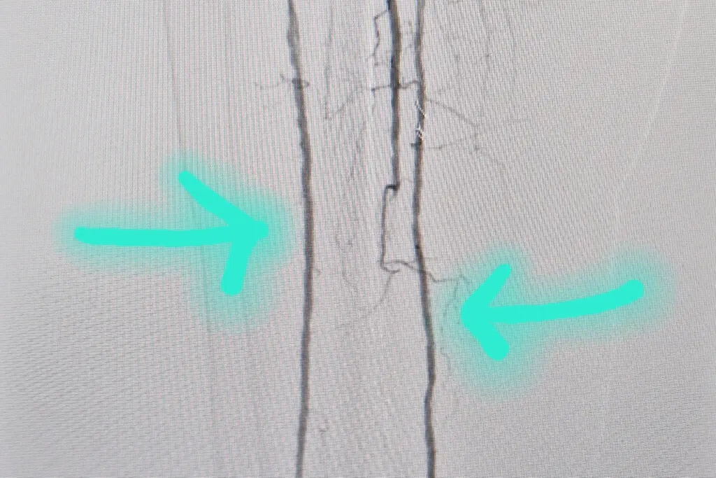
The arterial damage from diabetes affects the large arteries (atherosclerosis : section above on ‘Arterial Leg Ulcers) and small vessels as well (micro-angiopathy). Diabetic atherosclerosis in the legs is often located below the knee, which requires a Vascular and Endovascular Surgeon with expertise in distal(lower leg) bypass and crural (below knee) angioplasty. Atherosclerosis causes Peripheral Arterial Disease (PAD) which leads to poor healing and foot ulcers. Classically, PAD in diabetics damages the arteries below the knee (crural arteries).

Angiogram (X-ray picture of arteries using contrast or dye) of the blocked below knee arteries due to diabetic PAD. Gaps in the contrast indicate the site of blockage (red arrows)
Angiogram of diabetic patient after balloon angioplasty by Dr Yow & team. Green arrows indicate where angioplasty has restored in-line flow to the leg. This patient’s 6-month old leg ulcer healed within 2 weeks of the angioplasty’)
Ulcers in diabetics can be classified as pure neuropathic, pure arterial or mixed ulcers.
Diabetic foot ulcers are often located in the exposed or pressure points of the foot. This is where the foot is in contact with your shoe or exposed to minor trauma. Examples of these are on the balls of your feet, the heel, your big toes and little toes. Repeated, unnoticed abrasion or a cut from a nail/broken glass/bump against furniture will cause a wound. Poor blood supply due to atherosclerotic blockages results in a non-healing ulcer. Diabetic foot ulcers are often very deep and can go down to bone or tendon. Coupled with the diabetic’s poor ability to fight infection, this often leads to wet gangrene and limb or life-threatening diabetic foot sepsis.
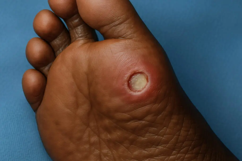
Diabetic foot ulcer in ball of foot with mild infection. See a Vascular Surgeon early.
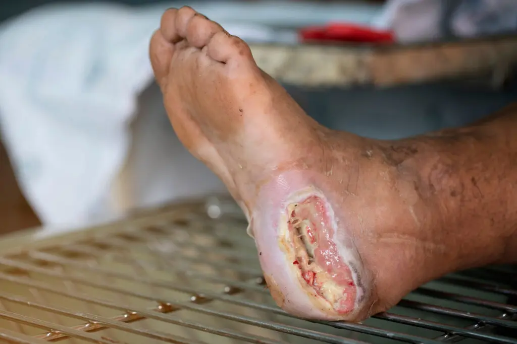
Neglected infected diabetic foot ulcer on pressure area of heel. Emergency treatment by a Vascular Surgeon can save the leg
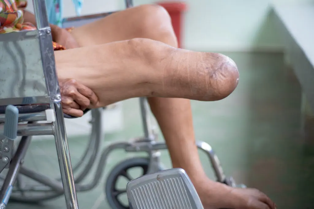
Amputations : We Can Prevent This!
It is crucial that you see a Vascular Surgeon immediately if you have a diabetic foot ulcer. DO NOT delay/have repeated washouts or dressings in a wound clinic without a Vascular review. Angioplasty often takes a couple of hours and is done under local anaesthesia. Bypass surgery takes a bit longer but the bypass graft stays open longer in the right hands. Hybrid operations combine the best of angioplasty/stenting and open surgery by reducing risk & physiological stress on the patient while offering the durabiliity of open surgery. Any one of these procedures can save your leg from further tissue loss or amputation.
RAPID ASSESSMENT AND TREATMENT BY ANGIOPLASTY/BYPASS/HYBRID VASCULAR INTERVENTION CAN SAVE YOUR LEG FROM AMPUTATION!
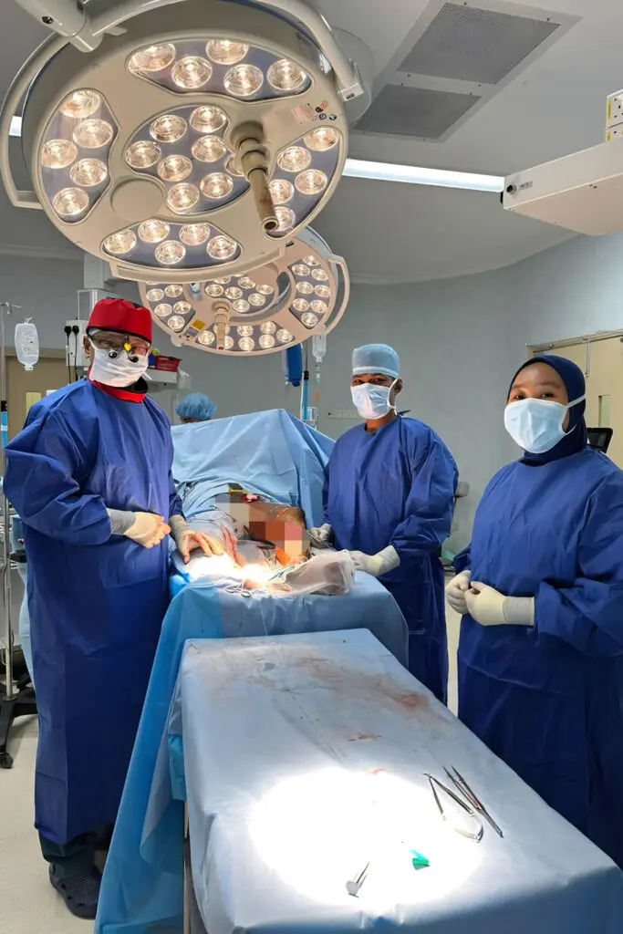
Mr Yow & his team performing a hybrid fem-distal bypass to save the leg of a diabetic patient
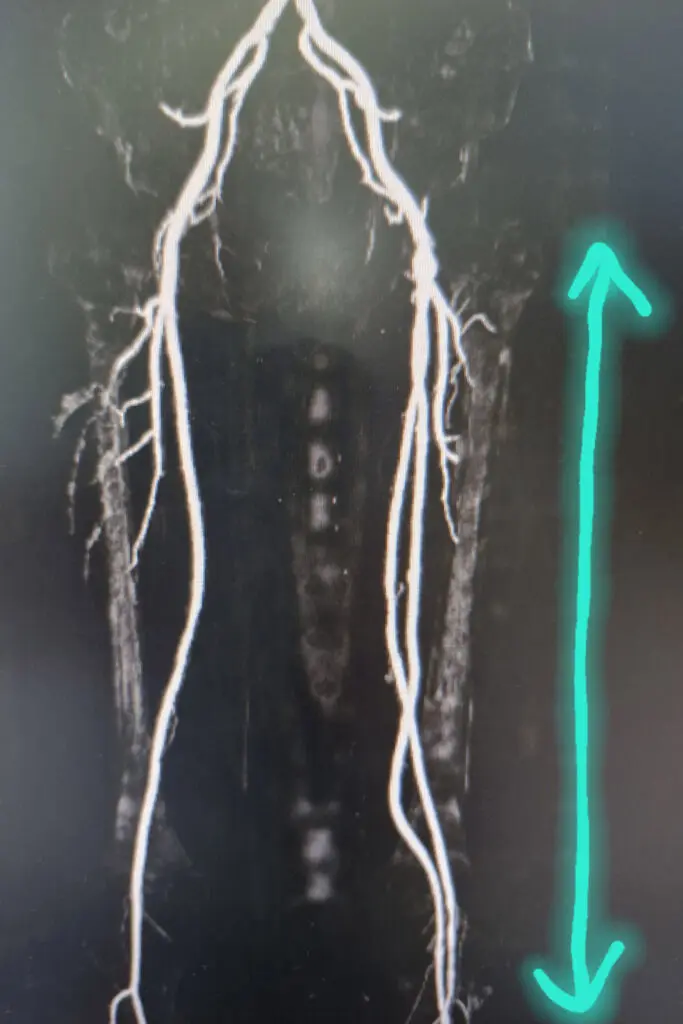
Post-operative CT angiogram after Mr Yow had removed clot by thrombectomy from the patient’s thigh & knee arteries and performed a vein bypass to the patient’s below knee (peroneal) artery
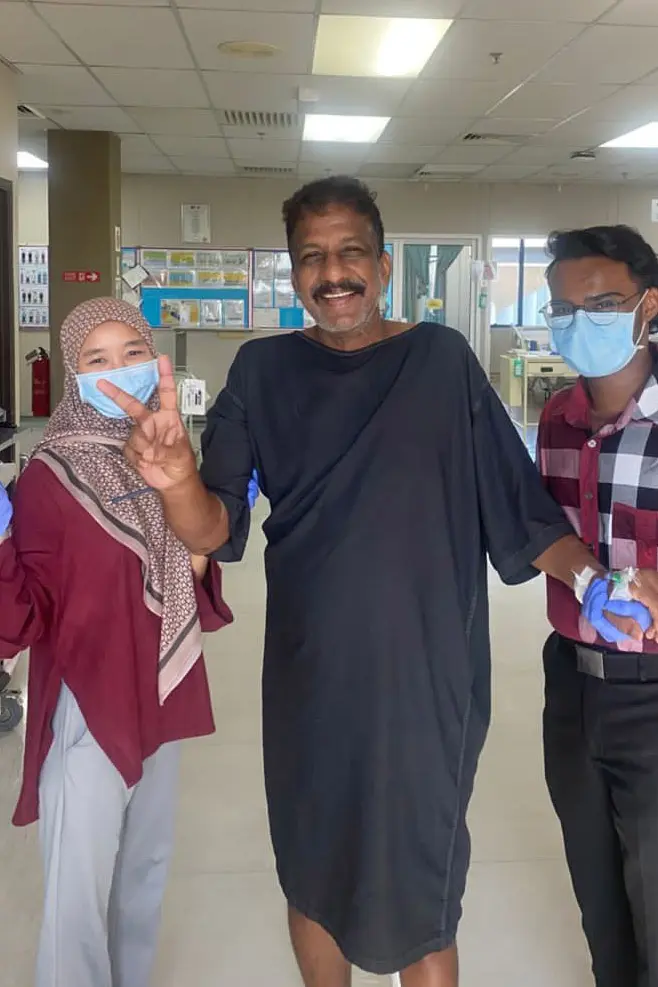
Diabetic patient after his successful revascularisation procedure walking on Day 2 after his operation. His numbness and pre-gangrene settled after surgery. Leg saved!